Periodontal Disease
The number one problem requiring oral health care is periodontal disease. Early intervention is the key and this
requires professional help. To properly evaluate an oral cavity and provide proper care the animal must be
anesthetized. It is impossible to deal with any problems in the back of the mouth or on the inside next to the
tongue in an awake animal. While ANY effort to keep the teeth clean is a plus, the reality is that anesthesia is
mandatory to perform a complete dental prophylaxis.
 |
 |
 |
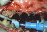
| Severe (Stage V) Periodontal Disease |
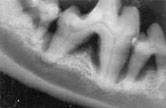
Xray Stage V Adult Periodontitis |
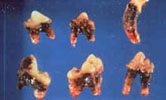
Hopeless Teeth |
Procedure for animal with minimal disease:
Under general anesthesia:
- Dental x-rays
- Supragingival (above the gumline) scaling
- Subgingival (below the gumline) scaling and curettage)
- Crown polishing
- Fluoride treatment
- Charting (record keeping)
The idea is to thoroughly clean above and below the gumline, smooth the enamel surface,
and improve the structure of the enamel layer. The time required will depend on the amount
of disease present. Animals with minimal disease may take up to 30 minutes to perform the
dental prophylaxis procedure. Animals with severe disease may take several hours of
anesthesia time and may require several visits to get the disease under control.
Unfortunately many animals hide their disease and the extent is not discovered until the animal is asleep.
Animals with advanced periodontal disease are another matter. In addition to the above procedure the following must be done:
- Surgical (open) curettage using flaps
- Extractions
- Perioceutic therapy (gels, solutions)
- Bone regeneration (bone morphogenic protein, synthetic materials)
- Antibiotic therapy
Advanced periodontal disease usually results from the effects long term dental plaque accumulation.
The implications for the animal's health can be severe in that severe periodontal disease can worsen
existing kidney or heart problems. The bacterial toxins absorbed into the body tax the animal's health
and create a feeling of general illness. Many animals that have their mouths returned to health
become active and playful again (giving their owners lots of guilt for not doing the procedure earlier!).
Periodontitis is a destructive inflammatory process of the peridontium driven by bacterial plaque
containing specific bacterial species that cause destruction of the gingiva, periodontal ligament,
alveolar bone and root cementum. In animals periodontitis is considered a progressive disease
with intermittent active episodes. In man gingivitis and periodontitis may be two separate diseases.
As periodontitis progresses the attachment mechanism is lost allowing deep tissue invasion of
microorganisms into the supporting structures resulting in periodontal ligament and alveolar bone loss.
In the final stages the tooth becomes mobile and is lost.
Of clinical significance is the type of bone loss occurring adjacent to the tooth root. Horizontal
bone loss is characterized by simple resorption of bone parallel to the cejs involving adjacent teeth
and remaining crestal bone supporting normal periodontium. (The
cej is a landmark on the tooth where the enamel stops and the
cementum [or root] starts, and if you draw a line connecting all the
cejs it will be almost a straight line. The plane of horizontal bone
loss is parallel to the cejs.) Vertical bone loss is characterized by
maintenance of crestal bone height with loss of periodontal ligament and supporting structure
adjacent to the tooth root. Horizontal bone loss usually has minimal "pocket formation" whereas
vertical bone loss has huge "pockets" requiring surgical intervention.
There are several established facts about periodontal microbiology. They are:
- Periodontal disease is caused by bacteria.
- Pockets cannot form in the absence of bacteria.
- Some of the pathogens have been identified; they are mostly gram negative anaerobic motile rods.
- The pathogens probably cause the disease.
The bacteria involved in periodontitis include:
- Porphyromonas gingivalis
- Prevotella species
- Fusobacterium species
- Bacteroides species
- Spirochetes
- Actinobacilus-actinomycetecomitans
On a percentage basis the number of aerobic bacteria decrease and anaerobic bacteria
increase as the disease progresses. When evaluating the microbiology in a diseased
mouth anaerobic cultures are indicated since aerobic cultures tend to be nonproductive.
Periodontitis can be characterized by the cardinal sign of destruction of the alveolar bone.
The features include conversion of junctional epithelium to pocket epithelium, and
destruction of the connective tissue matrix of gingiva and periodontal ligament.
Histologically periodontitis is characterized by several stages and closely
resembles a typical humoral immune response.
Initial lesion:
- Chronic vasculitis
- Excudation and invasion of neutrophils
- Junctional epithelial alterations
- Loss of perivascular structure (collagen)
Early lesion:
- Persistence of acute inflammation
- Predominance of lymphocytes
- Proliferation of junctional epithelium
- Loss of perivascular collagen
Established lesion:
- Persistence of acute inflammation
- Predominance of plasma cells
- Proliferation and migration of junctional epithelium
- Loss of collagen
- Presence of extra vascular immunioglobulins
Advanced lesion:
- Predominance of plasma cells
- Continued loss of collagen and fibrous connective tissue
- Pathologic alteration of plasma cells
- Loss of epithelial attachment
- Increased vascular supply
Most veterinary dentists refer to a staging system when characterizing an animal's mouth.
Because periodontitis is usually progressive, staging allows for improved record keeping
and treatment planning. None of the stages is totally unique and therefore a great deal
of variation can occur in an animal's mouth. The author uses a six stage system for adult
periodontitis. Stage 1 is gingivitis. Stage 2 is chronic gingivitis that does not progress to
periodontitis. Stages 3 through 6 are periodontitis characterized by attachment loss from
minimal (3) to maximum (6) and tooth loss. Obviously Stage 5 and 6 animals will require
aggressive "rocket science" therapy to maintain teeth while Stage 3 mouth will require
standard periodontal therapies.
Stage 1: acute gingivitis
- No attachment loss
- Limited to the gingival margin
- Neutrophil response
Stage 2: chronic gingivitis (greater than 6 mos)
- No attachment loss
- +/- stomatitis, buccal ulcers
- Lymphocytes/plasma cells predominate
Stage 3: early periodontitis
- Attachment loss
- Bone loss (1-3mm)
- No furcation involvement
- Lymphocytes/plasma cells present
Stage 4: established periodontitis
- Attachment loss
- Bone loss greater than 3mm
- Beginning furcation involvement (Classes 1 and 11)
- Lymphocytes, plasma cells predominate
Stage 5: advanced periodontitis
- Attachment loss
- Less than 1/3 root structure healthy
- Furcation involvement complete (Class 111)
- Plasma cells predominate
- Mobility (Grades 1and 2)
Stage 6: end stage periodontitis
- Attachment loss
- Bone loss to apex
- Plasma cell predominate
- Apical abscess present
- Combined perio/endo lesions
- Mobility (Grade 3)
Periodontitis can be localized or generalized. When generalized the staging system is
an average of the teeth involved (i.e., Stage 5) or a range (i.e., 3/5). When localized the specific teeth
are mentioned; i.e. #108 stage 5, # 208 stage 3. Regardless, record keeping of disease is
becoming the standard and if not done voluntarily the judicial system will dictate it.
The cornerstone of adult periodontitis therapy is the dental prophylaxis. The essence of therapy is to
control the subgingival infection via debridement of the root surface with or without flaps and with or
without antibiotics and to control plaque and reinfection. By definition a dental prophylaxis is the use
of appropriate procedures and/or techniques to prevent dental and oral disease and malformations.
The procedure includes:
- Supra and subgingival scaling
- Open/closed subgingival curettage
- Open/closed root planing
- Crown/root polishing
- Fluoride treatment
- Charting
 |
 |
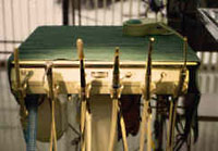
| Air-Driven Dental Handpieces |
Prophy Angle, Currette, Ultasonic Scaler |
Treatment planning for adult periodontitis is based on the stage of the disease. For generalized periodontitis the
following treatment plans are suggested as guidelines for therapy.
Stage 1:
- Dental prophylaxis
- Home care:
- Glyoxide
- CET toothpaste
- Maxiguard
- T/D diet
- Recall--6 to 12 months as needed
Stage 2:
- Dental prophylaxis
- Home care
- Chlorhexidine patches, rinses or gel pulse therapy
- Antibiotics--pulse therapy for life?
- Recall every 6 months
Stage 3:
- Dental prophylaxis with emphasis on subgingival cleaning; closed curettage and root
planing
- Home care
- Short term antibiotics
- Short term chlorhexidine patches, rinses or gel
- Long term Glyoxide or CET toothpaste
- Recall every 6 months
Stage 4:
- Pretreatment antibiotics ?
- Dental prophylaxis with emphasis on subgingival cleaning
- Open curettage/root planing (flaps)
- Home care
- Short term antibiotics
- Long term Glyoxide or CET toothpaste
- Recall every 3 to 6 months
Stage 5:
- Pretreatment with antibiotics
- Initial dental prophylaxis
- Open curettage/root planing (flaps)
- Extraction (hopeless teeth)
- Periodontal splinting mobile teeth
- Home care
- Antibiotics
- Twice a day oral hygiene
- AM-Glyoxide
- PM-Chlorhexidine
- Recall 2 to 4 weeks
- Follow up prophylaxis
- Repositioning flaps
- Guided tissue regeneration
- Recall every 3 months until stable then 6 months for life
Stage 6: Heroic therapy
- Pretreatment with antibiotics
- Initial dental prophylaxis
- Open curettage/root planing
- Periodontal splinting
- Extractions
- Home care (for life)
- Antibiotics
- Twice a day oral hygiene
- AM Glyoxide
- PM Chlorhexidine
- Recall 2 weeks
- Endodontic therapy
- Guided tissue regeneration
- Repositioning flaps
- Recall every 8 weeks until controlled
- Recall every 12 to 24 weeks for life
Obviously, the success of any treatment plan is based on the cooperation of the owner,
animal and veterinary dental team. Any breakdown in this equation spells failure.
Once the procedure for each visit is complete, accurate charting of the animal's mouth is imperative.
Without accurate record keeping, success or failure of treatment will be impossible to determine.
The Triadan Number System is computer compatible and is suggested as the system of choice.
Individual charts are based on personal preference. Small Animal Dentistry by Harvey and Emily
has several excellent dental charts for inspection.
Back to Main Treatments and Procedures Page
Return to Top of Page
|

